File:MastigiasMorphometrics.jpg
Original file (591 × 741 pixels, file size: 119 KB, MIME type: image/jpeg)
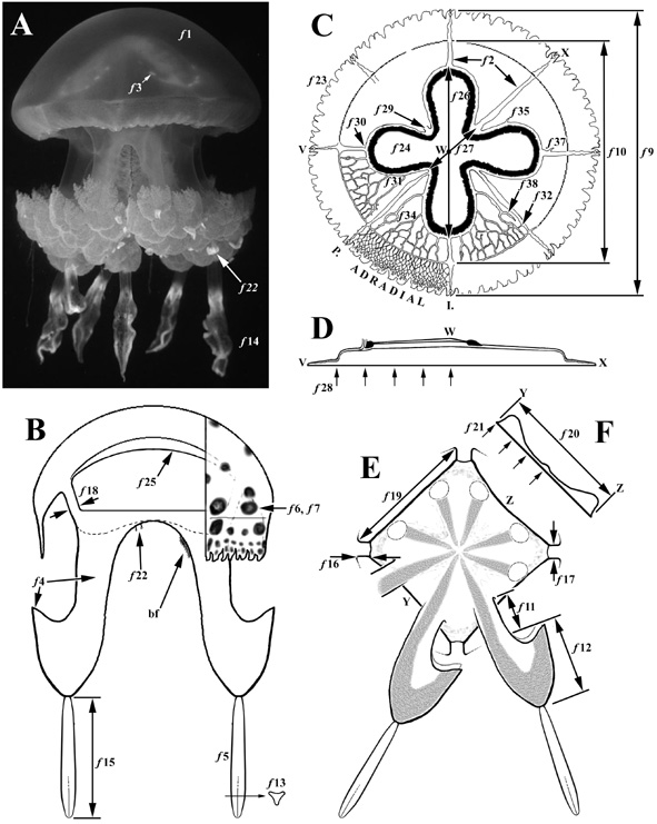
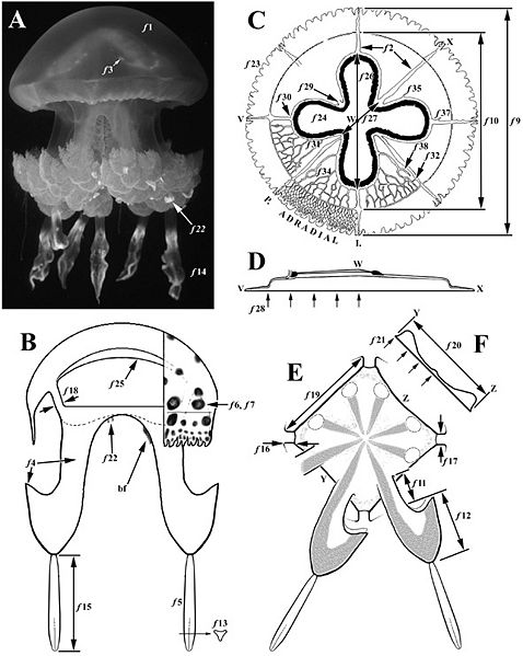
Figure: The morphology of Mastigias. (A) Photograph of an approximately 15 cm [bell diameter] medusa from Goby Lake, Palau. (B) Sketch of a longitudinal section along the interradial axis of the medusa [i.e. through the plane of the paper after rotation of the medusa in A through approximately 30° to the left]. (C) Sketch of the subumbrellar surface of the bell, and (D) cross-section through the bell along line VWX. (E) Schematic of the oral disc, in oral aspect, showing two of the eight oral arms, and (F) cross-section through the oral disc along line YZ or its orthogonal. Features discussed below include the following. Bell colour (f1). Perradial and interradial canal colour (f2). The presence of pigmented flecks at the roots or along the length of radial canals (f3). Oral arm colour (f4; lower arrow points to the �fringe�). Terminal club colour (f5). Abundance (f6) and colour (f7) of spots on the exumbrellar surface. Bell diameter (f9). Ring canal diameter (f10). Length of the simple, unwinged, portion of the oral arm (f11). Length of the winged portion of the oral arm (f12). Shape (f13), number (f14), and length (f15) of the terminal clubs. Length (f16), width (f17), and depth (f18) of the oral pillars. Width of the subgenital ostia (f19). Diameter (f20) and depth (f21) of the oral disc. Presence of intermediate filaments on the oral arm and oral disc (f22). Number of velar lappets [i.e. all lappets bar the two at each rhopalium] (f23). Shape of the gastrovascular cavity (f24). Colour of the subgenital porticus (f25). The interradial (I. f26) and perradial (P. f27) diameters of the gastrovascular cavity. Bell thickness (f28). The number of perradial (f29), interradial (f30), and adradial (f31) canal origins at the gastrovascular cavity. The number of perradial (f32) and adradial (f34) anastomoses in the radial canals that are circumscribed by the ring canal. The number of sinuses originating at the gastrovascular cavity (f35), and interradial (f37) and adradial (f38) canals. Radial canals are named consistent with the phylogenetic hypothesis of Uchida (1926: 87). Five features are not indicated in the figure: the mass of the whole medusa (f8), the number of interradial anastomoses (f33), the number of sinuses originating at perradial (f36) and ring canals (f39), and the number of anastomoses leading to two sinuses (f40). bf, brood filaments which are found only on mature female medusae.
File history
Click on a date/time to view the file as it appeared at that time.
| Date/Time | Thumbnail | Dimensions | User | Comment | |
|---|---|---|---|---|---|
| current | 02:46, 11 November 2009 | | 591 × 741 (119 KB) | Jshay (talk | contribs) | Figure: The morphology of Mastigias. (A) Photograph of an approximately 15 cm [bell diameter] medusa from Goby Lake, Palau. (B) Sketch of a longitudinal section along the interradial axis of the medusa [i.e. through the plane of the paper after rotation o |
You cannot overwrite this file.
File usage
The following page uses this file: